急性呼吸衰竭(acute respiratory failure, ARF)是重症肺炎死亡的重要原因,高流量氧疗(high-flow nasal cannula oxygen therapy, HFNC)和无创通气(non-invasive ventilation, NIV)等无创呼吸支持手段(non-invasive respiratory strategies, NIRS)已在早期广泛应用于这类患者,减少ARF患者对有创机械通气的需求。但若未及时识别治疗失败导致延迟插管,会使病死率明显升高[1]。因此,早期识别NIRS失败的患者非常重要。本研究的目的是建立并验证一种预测模型,用于评价超声参数在预测中度ARF患者NIRS治疗失败风险中的价值。
1 资料与方法 1.1 一般资料本研究为前瞻性观察性研究(试验注册号ChiCTR2000033162),筛选2020年10月至2023年1月收住苏州大学附属常熟医院重症医学科(intensive care unit, ICU)及急诊、呼吸病房未气管插管或气管切开的肺炎患者,纳入标准:100 mmHg≤氧合指数(PaO2/FiO2)≤200 mmHg,pH≥7.30(1 mmHg=0.133 kPa)。排除标准:年龄<18岁及妊娠者;慢性阻塞性肺病(chronic obstructive pulmonary disease, COPD)急性加重或哮喘持续状态;意识障碍;需要紧急插管、拒绝参与研究者。
NIRS措施包括:①HFNC(Fisher and Paykel Healthcar):初始设置气流速50 L/min,根据患者氧合调整FiO2,目标为氧饱和度(SpO2)>92%。;②NIV(Drager Savina 300或BELLAVISTA1000):初始设置:滴定驱动压力使潮气量维持在6~8 mL/kg(理想体重);以SpO2>92%为目标滴定PEEP及FiO2;每日实施8 h以上。因既往文献及本课题组前期研究显示HFNC及NIV在治疗效果相似[2-3],因此将两种呼吸支持措施合并分析。
治疗失败定义为治疗期间患者需要转为有创通气或死亡。需要转为有创通气指征[4]:至少满足以下两条:①呼吸急促>35次/min;②pH<7.25;③SpO2<88%持续5 min以上;④格拉斯哥昏迷评分<12分。本研究通过苏州大学附属常熟医院医学伦理委员会的审核(2020伦审(申报)批第12号),并获得患者或授权委托人知情同意。
1.2 监测指标记录患者入院时基本情况,包括年龄、性别、基础疾病等,入院及治疗24 h时生命体征、血气分析指标、ROX指数[SPO2/(FIO2·RR)]、肺部超声评分(lung ultrasound score, LUS)、膈肌移动度(diaphragmatic excursion, DE)、舒张末期右心室与左心室比值(right venticular/left venticular end-diastolic area, RV/LV),及28 d病死率、滞留ICU时间。
1.3 统计学方法将纳入研究的患者随机(7∶3)分为建模组和验证组。用(x±s)描述连续变量符合正态分布的数据,中位数(四分位间距)描述非正态分布的数据,正态分布数据组间比较使用成组t检验,非分布数据使用Mann-Whitney U秩和检验,率的比较用χ2检验。计算容忍度检验自变量是否存在多重共线性,并对单因素分析差异有统计学意义的因素进行多因素Logistic回归分析。利用建模组中的数据构建预测模型,并在验证组中进行验证。通过ROC曲线评价模型的诊断效果,绘制校准曲线评价一致性,计算净重新分类指数(net reclassification index, NRI)评价模型的预测能力,决策曲线分析(decision curve analysis,DCA)评价净效益。最后使用R语言绘制预测模型的列线图。所有统计分析均采用SPSS 22.0及R 3.0.1语言进行。以P<0.05为差异有统计学意义。
2 结果 2.1 基本情况2020年10月至2023月1月ICU及急诊、呼吸病房共收治急性呼吸衰竭患者242例,其中COPD急性加重或哮喘持续状态34例,意识障碍5例,需要紧急插管、拒绝参与研究3例,中途放弃治疗7例予以排除,最终193例患者纳入研究。其中138例(71.5%)初始治疗为HFNC,55例(28.5%)为NIV。共112例(58%)NIRS失败,其中HFNC 81例(58.7%),NIV31例(56.4%)(χ2=0.088,P=0.767)。54例(28%)在入院后28 d内死亡,其中HFNC 37例(26.8%),NIV 17例(30.9%),(χ2=0.328,P=0.567)。将患者按7:3随机分为建模组137例及验证组56例,两组患者基本情况差异无统计学意义,见表 1。
| 临床特征 | 建模组(n=137) | 验证组(n=56) | 检验值(Z/t/χ2) | P值 |
| 男性(n,%) | 99(72.3) | 42(75) | 0.151 | 0.697 |
| 年龄[岁,(x±s)] | 72.1±9.1 | 70.8±9.6 | 0.881 | 0.380 |
| 基础疾病(n,%) | ||||
| 高血压 | 64(46.7) | 32(57.1) | 1.729 | 0.189 |
| 糖尿病 | 22(16.1) | 9(16.1) | 0.000 | 0.998 |
| 哮喘或COPD | 32(23.4) | 13(23.2) | 0.000 | 0.983 |
| 慢性肝病 | 6(4.4) | 4(7.1) | 0.183 | 0.668 |
| 慢性肾功能不全 | 17(12.4) | 7(12.5) | 0.000 | 0.986 |
| pH (x±s) | 7.41±0.04 | 7.42±0.04 | -1.685 | 0.094 |
| PaO2/FiO2 [mmHg, M(Q1, Q3)] | 166.0(148.5, 179.0) | 168.0(150.0, 179.8) | -0.763 | 0.446 |
| PaCO2[mmHg, (x±s)] | 36.1±3.6 | 35.7±3.1 | 0.608 | 0.544 |
| RR[次/min,(x±s)] | 29.5±8.0 | 28.8±8.1 | 0.528 | 0.598 |
| HR[次/min,M(Q1, Q3)] | 80.0(74.5, 85.0) | 78.0(73.3, 82.0) | -1.640 | 0.101 |
| MAP[mmHg, (x±s)] | 78.9±7.2 | 78.6±6.7 | 0.240 | 0.810 |
| ROX指数[M(Q1, Q3)] | 5.40(4.45, 6.80) | 5.80(4.25, 6.50) | -0.615 | 0.539 |
| LUS[分, M(Q1, Q3)] | 12.0(9.0, 14.0) | 12.0(10.0, 15.0) | -1.290 | 0.197 |
| DE[cm, M(Q1, Q3)] | 1.20(0.90, 1.62) | 1.30(1.08, 1.69) | -1.096 | 0.273 |
| RV/LV[%, x±s] | 69.2±14.2 | 69.6±16.4 | -0.200 | 0.842 |
| 初始治疗措施(n,%) | ||||
| HFNC | 99(72.3) | 39(69.6) | 0.134 | 0.741 |
| NIV | 38(27.7) | 17(30.4) | 0.134 | 0.741 |
| 有创机械通气(n,%) | 84(61.3) | 28(50.0) | 2.089 | 0.148 |
| 有创机械通气时间[d, M(Q1, Q3)] | 11.0(5.0~16.0) (n=84) | 11.5(4.3, 14.8) (n=28) | -0.495 | 0.621 |
| 血管活性药物的使用(n,%) | 56(40.9) | 22(39.3) | 0.042 | 0.838 |
| 28 d病死率(n,%) | 39(28.5) | 15(26.8) | 0.056 | 0.813 |
| 住院时间[d, M(Q1, Q3)] | 17.0(12.0~25.0) | 17.0(11.0-24.8) | -0.068 | 0.946 |
| 注:COPD为慢性阻塞性肺病,RR(respiratory rate)为呼吸频率,HR(heart rate)为心率,MAP(mean arterial pressure)为平均动脉压,LUS(lung ultrasound score)为肺部超声评分,DE(diaphragmatic excursion)为膈肌移动度。RV/LV((right venticular/left venticular end-diastolic area)为舒张末期右心室与左心室比值 | ||||
建模组NIRS失败组患者入院时及24 h PaO2/FiO2、DE均低于成功组(P均 < 0.01),RV/LV均高于成功组(P=0.013, < 0.01),24 h RR及LUS评分高于成功组而ROX指数低于成功组(P均 < 0.01),此外失败组血管活性药物的使用率升高(P=0.001),差异有统计学意义(见表 2)。选择单因素分析统计学差异更明显的24 h时各指标纳入多因素Logistic回归分析,各自变量经线性回归检验不存在多重共线性,方差膨胀因子分别为血管活性药物使用率1.025,PaO2/FiO2 1.105,RR 5.753,ROX指数5.605,LUS评分1.031,DE 1.129,RV/LV 1.098,结果显示NIRS失败的独立危险因素是血管活性药物使用(OR=4.709, P=0.012)、较高的RR(OR=1.254, P=0.035)、LUS评分(OR=1.250, P=0.037)及RV/LV(OR=1.057, P=0.008),而PaO2/FiO2(OR=0.950, P=0.001)、DE为保护因素(OR=0.107, P=0.001)(见表 3)。
| 指标 | 成功组(n=53) | 失败组(n=84) | 检验值(Z/t/χ2) | P值 |
| 男性(n,%) | 35(66.0) | 64(76.2) | 1.671 | 0.196 |
| 年龄[岁,(x±s)] | 70.7±9.1 | 73.0±8.9 | -1.475 | 0.143 |
| 基础疾病(n,%) | ||||
| 高血压 | 23(43.4) | 41(48.8) | 0.383 | 0.536 |
| 糖尿病 | 6(11.3) | 16(19.0) | 1.439 | 0.230 |
| 哮喘或COPD | 10(18.9) | 22(26.2) | 0.973 | 0.324 |
| 慢性肝病 | 2(3.8) | 4(4.8) | 0.077 | 0.781 |
| 慢性肾病 | 7(13.2) | 10(11.9) | 0.051 | 0.822 |
| 初始治疗方式(HFNC)(n,%) | 40(75.5) | 59(70.2) | 0.444 | 0.505 |
| 血管活动药物使用 | 12(22.6) | 44(52.4) | 11.892 | 0.001 |
| pH (x±s) | ||||
| 0 h | 7.41±0.04 | 7.41±0.04 | -0.454 | 0.651 |
| 24 h | 7.41±0.03 | 7.42±0.03 | -1.125 | 0.262 |
| PaO2/FiO2 [mmHg, M(Q1-Q3)] | ||||
| 0 h | 178.0(167.0, 187.0) | 155.0(134.0, 168.8) | -5.878 | < 0.01 |
| 24 h | 178(169.5, 188.0) | 162.0(140.8, 176.8) | -5.201 | < 0.01 |
| PaCO2 [mmHg, (x±s)] | ||||
| 0 h | 35.7±3.3 | 36.3±3.7 | -0.857 | 0.393 |
| 24 h | 35.0±2.7 | 35.9±4.5 | -1.379 | 0.170 |
| RR [次/min, M(Q1-Q3)] | ||||
| 0 h | 28.0(22.0, 33.0) | 30.0(24.0, 35.0) | -1.161 | 0.245 |
| 24 h | 26.0(21.0, 31.5) | 34.0(29.0, 39.0) | -5.244 | < 0.01 |
| HR [次/min, (x±s)] | ||||
| 0 h | 79.5±9.1 | 80.3±7.7 | -0.511 | 0.610 |
| 24 h | 78.0±10.3 | 78.9±7.9 | -0.577 | 0.565 |
| MAP[mmHg, (x±s)] | ||||
| 0 h | 78.5±7.3 | 79.1±7.2 | -0.496 | 0.621 |
| 24 h | 76.9±7.3 | 79.5±8.0 | -1.907 | 0.059 |
| ROX指数[M(Q1, Q3)] | ||||
| 0 h | 5.60(4.80, 7.10) | 5.20(4.40, 6.60) | -1.652 | 0.099 |
| 24 h | 6.10(5.10, 7.40) | 4.59(4.10, 5.68) | -5.337 | < 0.01 |
| LUS [分, M(Q1, Q3)] | ||||
| 0 h | 12.0(9.0, 13.0) | 12.0(9.0, 15.0) | -1.481 | 0.139 |
| 24 h | 12.0(10.0, 13.0) | 13.0(11.0, 15.0) | -2.818 | 0.005 |
| DE [cm, M(Q1, Q3)] | ||||
| 0 h | 1.40(1.13, 1.74) | 1.07(0.88, 1.46) | -3.174 | 0.002 |
| 24 h | 1.66(1.29, 1.89) | 1.07(0.84, 1.46) | -5.535 | < 0.01 |
| RV/LV [%, (x±s)] | ||||
| 0 h | 65.4±14.9 | 71.6±13.3 | -2.529 | 0.013 |
| 24 h | 63.2±15.5 | 72.2±12.4 | -3.767 | < 0.01 |
| 28 d病死率(n,%) | ||||
| 住院时间[d, M(Q1, Q3)] | ||||
| 注:COPD为慢性阻塞性肺病,RR(respiratory rate)为呼吸频率,HR(heart rate)为心率,MAP(mean arterial pressure)为平均动脉压,LUS(lung ultrasound score)为肺部超声评分,DE(diaphragmatic excursion)为膈肌移动度。RV/LV((right venticular/left venticular end-diastolic area)为舒张末期右心室与左心室比值 | ||||
| 参数 | B | S.E. | Wald | P值 | OR | 95%CI |
| 血管活性药物 | 1.550 | 0.615 | 6.358 | 0.012 | 4.709 | 1.412~15.705 |
| PaO2/FiO2 | -0.051 | 0.016 | 10.431 | 0.001 | 0.950 | 0.921~0.980 |
| RR | 0.226 | 0.107 | 4.447 | 0.035 | 1.254 | 1.016~1.548 |
| LUS | 0.223 | 0.107 | 4.336 | 0.037 | 1.250 | 1.013~1.541 |
| DE | -2.233 | 0.667 | 11.211 | 0.001 | 0.107 | 0.029~0.396 |
| RV/LV | 0.056 | 0.021 | 6.925 | 0.008 | 1.057 | 1.014~1.102 |
| 常数 | -3.485 | 6.165 | 0.319 | 0.572 | 0.031 | |
| 注:RR(respiratory rate)为呼吸频率,LUS(lung ultrasound score)为肺部超声评分,DE(diaphragmatic excursion)为膈肌移动度。RV/LV(right venticular/left venticular end-diastolic area)为舒张末期右心室与左心室比值 | ||||||
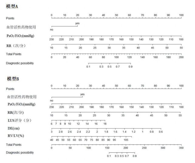
基于多因素分析中差异有统计学意义的各指标,构建预测NIRS失败的模型,以不含超声参数的临床指标构建模型A,超声参数联合临床指标构建模型B,模型B的AIC值小于模型A(表 4)。绘制各模型ROC曲线,建模组模型B的曲线下面积(AUC)大于模型A,差异有统计学意义(0.872 vs. 0.928, P=0.009);验证组中模型A及模型B的曲线下面积分别为0.867, 0.932(P=0.07)(表 5,图 1)。校准图显示在建模组及验证组中两个模型校准度均较好(P均 > 0.05)(图 2)。NRI指数分析显示与模型A相比,模型B预测能力更强(NRI=0.758,P < 0.01),在总人群中正确分类比例提高75.8%,在治疗失败组中提高38.1%,成功组中提高37.7%(表 6)。DCA曲线显示当预测值小于80%时,模型B的净效益优于模型A(图 3)。
| 参数 | B | OR | 95%CI | AIC |
| 模型A | 125.57 | |||
| 血管活性药物 | 1.374 | 3.952 | 1.534~10.950 | |
| PaO2/FiO2 | -0.053 | 0.948 | 0.923~0.970 | |
| RR | 0.255 | 1.291 | 1.101~1.556 | |
| 常数 | -1.124 | |||
| 模型B | 103.77 | |||
| 血管活性药物 | 1.550 | 4.709 | 1.412~15.705 | |
| PaO2/FiO2 | -0.051 | 0.950 | 0.921~0.980 | |
| RR | 0.226 | 1.254 | 1.016~1.548 | |
| LUS | 0.223 | 1.250 | 1.013~1.541 | |
| DE | -2.233 | 0.107 | 0.029~0.396 | |
| RV/LV | 0.056 | 1.057 | 1.014~1.102 | |
| 常数 | -3.485 | 0.031 | ||
| 注:RR(respiratory rate)为呼吸频率,LUS(lung ultrasound score)为肺部超声评分,DE(diaphragmatic excursion)为膈肌移动度。RV/LV(right venticular/left venticular end-diastolic area)为舒张末期右心室与左心室比值 | ||||
| 分组 | 模型 | AUC | Cut-off值 | 特异度(%) | 敏感度(%) | P值 (与模型B相比) |
| 建模组 | A | 0.872 | 0.641 | 88.7 | 77.4 | 0.009 |
| B | 0.928 | 0.578 | 92.5 | 85.7 | - | |
| 验证组 | A | 0.867 | 0.667 | 89.3 | 67.9 | 0.07 |
| B | 0.932 | 0.689 | 89.3 | 85.7 | - |

|
| 图 1 各预测模型的ROC曲线 Fig 1 ROC curve for each prediction model |
|
|

|
| A:建模组模型A; B验证组模型A; C:建模组模型B; D:验证组模型B 图 2 各预测模型的校准图 Fig 2 Calibration plot for each prediction model |
|
|
| 组别 | NRI | 95%CI | Z | P值 |
| 总人群 | 0.758 | 0.440~1.088 | 4.547 | < 0.01 |
| 治疗失败组 | 0.381 | 0.181~0.506 | 4.398 | < 0.01 |
| 治疗成功组 | 0.377 | 0.208~0.635 | 3.498 | < 0.01 |

|
| 红色曲线:模型A,蓝色曲线:模型B,灰色曲线:全部获益;黑色曲线:均不获益 图 3 各预测模型的DCA曲线 Fig 3 Decision curve analysis (DCA) for each prediction model |
|
|

|
| RR(respiratory rate)为呼吸频率,LUS(lung ultrasound score)为肺部超声评分,DE(diaphragmatic excursion)为膈肌移动度。RV/LV((right venticular/left venticular end-diastolic area)为舒张末期右心室与左心室比值 图 4 各预测模型的列线图 Fig 4 Nomograms for each prediction model |
|
|
绘制预测NIRS失败的模型列线图。通过各因素对应的顶部积分表计算所对应的分值,将各项分值相加得到总分,再根据底部总分值表计算预测NIRS失败的概率。
3 讨论ARF是ICU中最常见的疾病之一。COVID-19流行前的多中心调查就显示ICU中ARF发生率达13%[5],在COVID-19流行期间,ARF的发生率更是明显升高,感染患者中有49.5%出现急性呼吸窘迫综合征(acute respiratory distress syndrome, ARDS)[6],即使是在青少年患者中,也有约1/3的患者出现ARF[7]。近5年来美国卫生统计中心的数据显示ARF相关的病死率每年增加约3.4%,与ARDS相关的病死率在35%~45%之间,近几年未有下降[8-9]。本研究中患者的28 d病死率为28%,病死率略低于其他研究的原因可能与本研究对象主要以中度急性呼吸衰竭患者有关。
NIRS可在促进气体交换的同时保留自主意识及自主呼吸,在COVID-19引起的ARF患者中,有41%的患者接受了NIRS,与传统氧疗相比,NIRS在降低病死率和气管插管风险方面有重要作用[10]。但其在PaO2/FiO2≤200 mmHg患者中仍有着不低的失败率,本研究中58%的患者NIRS失败,其中HFNC 81例(58.7%)、NIV31例(56.4%),符合之前报道的HFNC失败率在28%~66%、NIV 11%~79%之间。在不同研究中失败率的差异较大,可能与参数的设置、使用时间及患者依从性等因素相关[11]。
由于使用NIRS的患者生理氧储备较少,一旦延迟插管就可能导致不良后果。NIRS失败已被发现是ARF患者死亡的独立危险因素[12]。因此,临床上需权衡避免插管的益处与延迟插管的危害性,早期预测NIRS失败可以帮助临床医生适当分配危重症资源。由于既往及前期研究发现HFNC和NIV临床疗效上相似[2-3],因此本研究未再将HFNC和NIV进行分组分析。
本研究结果显示在临床指标中PaO2 /FiO2、RR及血管活性药物的使用预测NIRS失败的鉴别能力较高。PaO2 /FIO2、RR是临床上最常用于判断氧合情况及呼吸疲劳的指标。既往研究有通过使用HACOR预测量表(HR、pH、格拉斯哥昏迷量表、PaO2 /FiO2、RR)进行预测NIV失败及病死率,24 h HACOR评分 > 5与病死率增加相关(RR 2.39, 95% CI 1.60~3.56)[13]。也有研究使用此评分对非COVID~19患者NIRS失败进行预测,其C统计量在0.83(95%CI 0.81~0.87),与本研究中的模型A相似[14]。在COVID-19流行期间由于医疗资源有限,故有研究采用ROX指数预测HFNC失败的风险,但其在使用上仍存在局限性。首先,SpO2的上限为100%,与PaO2也并非呈线性关系,有无法识别呼吸恶化的风险;此外,在危重症患者中较常可见PaCO2值下降,导致解离曲线向左偏移,使测得的SpO2值高估实际氧合状态。且目前研究对于ROX阈值在不同类型患者中并不统一,从3.67到5.37不等,临床使用上价值有限[15-16]。针对ICU收治的ARF患者,PaO2是常规需要监测的指标,因此如本研究结果所示,相比ROX指数,PaO2/FiO2的预测价值更高。有研究通过使用PaO2改良的mROX指数[PaO2/(FiO2·RR)]用于早期预测HFNC治疗是否成功,结果显示2 h时mROX为4.3时敏感性96.1%,特异性70.8%,其ROC的曲线下面积高于ROX指数,分别为0.952 vs. 0.911(COVID-19患者),0.806 vs. 0.756(非COVID-19肺炎患者)[17]。本研究中计算mROX指数,其预测NIRS失败的的阈值在入院时为4.9108,灵敏度81.1%, 特异度50.0%,24 h的阈值为5.834,灵敏度77.4%, 特异度80.0%。此外,笔者也发现使用大剂量血管活动药物是NIRS失败的独立危险因素,但鉴别能力偏低。与之前的研究结果一致[14]。
ARF患者氧合及炎症情况与肺部影像学的表现密切相关,肺部超声可以使用B线来识别肺部炎症及渗出,操作简便、无辐射,可重复。在对柏林ARDS定义的修改中,肺超声也被提议作为胸片的替代方案[18]。LUS是监测ARDS肺通气变化的可靠床边工具,其总评分及腹侧、中间及背侧区域的评分与CT计算的通气量显著相关(分别为r= - 0.74、- 0.66、- 0.69、- 0.63;P < 0.05)[19]。膈肌功能障碍是呼吸衰竭的最早信号之一。在接受HFNC治疗的患者中,有48%的患者出现膈肌收缩减弱,13%患者出现膈肌收缩异常[20]。Tonelli等证实了超声测量膈肌收缩速度可作为预测HFNC成功的指标,治疗12 h后仰卧膈肌收缩速度 > 1.35 cm/s预测成功的敏感性为89%,特异性为57%[21]。此外,潮气量与膈肌之间还存在线性相关关系,因此膈肌超声可能成为在早期阶段预测ARF治疗效果的工具[22]。在ARF患者中由于肺循环压力升高,右心室难以适应压力负荷的增加,导致右心室功能障碍。43%的中重度ARDS机械通气患者出现急性右心室增大,15%出现肺动脉高压[23]。对仅接受有创机械通气的COVID-19患者进行RV-散斑跟踪超声心动图应变分析显示28.7%的患者RV自由壁纵向应变异常,且其与30 d病死率显著相关(OR 2.22,95% CI 1.14~4.39,P= 0.020)[24]。其他研究也表明新发RV衰竭是ARDS患者中90 d病死率独立相关的因素(调整后HR 8.17,95%CI 3.15~21.2,P < 0.001)。PaO2/FiO2、PaCO2、通气比和驱动压力显著恶化,这些都可能是导致RV衰竭的原因[25]。ARF患者中关注预防右心室功能障碍,针对RVF及时处理,可能对这些患者的预后有益处。但是在ARF的早期阶段,关于超声评估的临床数据非常缺乏,大多数研究都集中在撤机阶段[26]。Kundu等[27]曾采用联合肺、膈肌和心脏超声的方案预测拔管失败的价值,失败组患者在自主呼吸试验期间膈肌增厚分数 < 26%(OR 6.20, 95% CI 1.06~36.04),速度时间积分对被动抬腿的反应变化 < 10.2%(OR 6.16, 95% CI 1.14~33.13)是撤机失败的预测因素。本研究分别从肺部炎症、膈肌功能及右心功能三方面选取超声指标,联合呼吸频率、氧合指数等临床关参数建立了模型B,结果显示其在预测NIRS失败方面的鉴别能力,优于仅基于临床参数的模型A。
本研究仍有局限性。首先本研究为单中心研究,结果仍需在其他中心进行验证。其次,纳入的患者中涵盖了2022年12月至2023年2月入院的COVID-19感染患者,由于当时病例激增,可能导致在治疗措施选择上和平时有所不同。第三,没有测量超声操作研究人员间或研究人员内的变异性。
利益冲突 所有作者声明无利益冲突
作者贡献声明 俞隼、许春阳:直接参与研究设计、数据收集分析、论文撰写;叶宏伟:研究设计、文章审阅与指导;黄晟:数据收集整理分析、文章内容修改
| [1] | Via G, Vasques F, Hussain A, et al. Bedside noninvasive monitoring of mechanically ventilated patients[J]. Curr Opin Crit Care, 2021, 27(1): 66-75. DOI:10.1097/MCC.0000000000000789 |
| [2] | Yasuda H, Okano H, Mayumi T, et al. Association of noninvasive respiratory support with mortality and intubation rates in acute respiratory failure: a systematic review and network meta-analysis[J]. J Intensive Care, 2021, 9(1): 32. DOI:10.1186/s40560-021-00539-7 |
| [3] | 俞隼, 许春阳, 叶宏伟, 等. 高流量氧疗在中度急性呼吸衰竭患者中的应用价值[J]. 中华急诊医学杂志, 2022, 31(9): 1236-1242. DOI:10.3760/cma.j.issn.1671-0282.2022.09.014 |
| [4] | Frat JP, Thille AW, Mercat A, et al. High-flow oxygen through nasal cannula in acute hypoxemic respiratory failure[J]. N Engl J Med, 2015, 372(23): 2185-2196. DOI:10.1056/NEJMoa1503326 |
| [5] | Villar J, Mora-Ordoñez JM, Soler JA, et al. The PANDORA study: prevalence and outcome of acute hypoxemic respiratory failure in the pre-COVID-19 era[J]. Crit Care Explor, 2022, 4(5): e0684. DOI:10.1097/CCE.0000000000000684 |
| [6] | Wang FL, Ran LY, Qian CC, et al. Epidemiology and outcomes of acute kidney injury in COVID-19 patients with acute respiratory distress syndrome: a multicenter retrospective study[J]. Blood Purif, 2021, 50(4/5): 499-505. DOI:10.1159/000512371 |
| [7] | Kest H, Kaushik A, Shaheen S, et al. Epidemiologic characteristics of adolescents with COVID-19 disease with acute hypoxemic respiratory failure[J]. Crit Care Res Pract, 2022, 2022: 7601185. DOI:10.1155/2022/7601185 |
| [8] | Cui YQ, Ding XF, Liang HY, et al. Efficacy and safety of low-dose corticosteroids for acute respiratory distress syndrome: a systematic review and meta-analysis[J]. World J Emerg Med, 2021, 12(3): 207-213. DOI:10.5847/wjem.j.1920-8642.2021.03.008 |
| [9] | Meyer NJ, Gattinoni L, Calfee CS. Acute respiratory distress syndrome[J]. Lancet, 2021, 398(10300): 622-637. DOI:10.1016/S0140-6736(21)00439-6 |
| [10] | Menga LS, Berardi C, Ruggiero E, et al. Noninvasive respiratory support for acute respiratory failure due to COVID-19[J]. Curr Opin Crit Care, 2021, 28(1): 25-50. DOI:10.1097/mcc.0000000000000902 |
| [11] | Bellani G, Laffey JG, Pham T, et al. Noninvasive ventilation of patients with acute respiratory distress syndrome. insights from the LUNG SAFE study[J]. Am J Respir Crit Care Med, 2017, 195(1): 67-77. DOI:10.1164/rccm.201606-1306OC |
| [12] | Munshi L, Mancebo J, Brochard LJ. Noninvasive respiratory support for adults with acute respiratory failure[J]. N Engl J Med, 2022, 387(18): 1688-1698. DOI:10.1056/NEJMra2204556 |
| [13] | Innocenti F, Giordano L, Gualtieri S, et al. Prediction of mortality with the use of noninvasive ventilation for acute respiratory failure[J]. Respir Care, 2020, 65(12): 1847-1856. DOI:10.4187/respcare.07464 |
| [14] | Liu L, Xie JF, Wu WJ, et al. A simple nomogram for predicting failure of non-invasive respiratory strategies in adults with COVID-19: a retrospective multicentre study[J]. Lancet Digit Health, 2021, 3(3): e166-e174. DOI:10.1016/S2589-7500(20)30316-2 |
| [15] | Ferrer S, Sancho J, Bocigas I, et al. ROX index as predictor of high flow nasal cannula therapy success in acute respiratory failure due to SARS-CoV-2[J]. Respir Med, 2021, 189: 106638. DOI:10.1016/j.rmed.2021.106638 |
| [16] | Maraziti G, Becattini C. Early variation of respiratory indexes to predict death or ICU admission in severe acute respiratory syndrome coronavirus-2-related respiratory failure[J]. Respiration, 2022, 101(7): 632-637. DOI:10.1159/000522275 |
| [17] | Li Z, Chen C, Tan ZJ, et al. Prediction of high-flow nasal cannula outcomes at the early phase using the modified respiratory rate oxygenation index[J]. BMC Pulm Med, 2022, 22(1): 227. DOI:10.1186/s12890-022-02017-8 |
| [18] | Ranieri VM, Rubenfeld G, Slutsky AS. Rethinking acute respiratory distress syndrome after COVID-19: if a "better" definition is the answer, what is the question?[J]. Am J Respir Crit Care Med, 2023, 207(3): 255-260. DOI:10.1164/rccm.202206-1048CP |
| [19] | Andrea C, Irene S, Emanuele P, et al. Clinical performance of lung ultrasound in predicting time-dependent changes in lung aeration in ARDS patients[J]. J Clin Monit Comput, 2023, 37(2): 473-480. DOI:10.1007/s10877-022-00902-5 |
| [20] | Takashima T, Nakanishi N, Arai Y, et al. The effect of high-flow nasal cannula on diaphragm dysfunction including paradoxical diaphragmatic contraction in the intensive care unit[J]. J Med Invest, 2021, 68(1.2): 159-164. DOI:10.2152/jmi.68.159 |
| [21] | Tonelli R, Fantini R, Tabbì L, et al. Early inspiratory effort assessment by esophageal manometry predicts noninvasive ventilation outcome in De novo respiratory failure. A pilot study[J]. Am J Respir Crit Care Med, 2020, 202(4): 558-567. DOI:10.1164/rccm.201912-2512OC |
| [22] | Bruna M, Hidalgo G, Castañeda S, et al. Diaphragmatic ultrasound predictors of high-flow nasal Cannula therapeutic failure in critically ill patients with SARS-CoV-2 pneumonia[J]. J Ultrasound Med, 2023, 42(6): 1277-1284. DOI:10.1002/jum.16141 |
| [23] | 张磊, 陈国忠, 王华英, 等. 膈肌超声评估机械通气撤机中临床应用价值[J]. 中华急诊医学杂志, 2023, 32(2): 241-246. DOI:10.3760/cma.j.issn.1671-0282.2023.02.020 |
| [24] | McErlane J, McCall P, Willder J, et al. Right ventricular free wall longitudinal strain is independently associated with mortality in mechanically ventilated patients with COVID-19[J]. Ann Intensive Care, 2022, 12(1): 104. DOI:10.1186/s13613-022-01077-7 |
| [25] | Evrard B, Goudelin M, Giraudeau B, et al. Right ventricular failure is strongly associated with mortality in patients with moderate-to-severe COVID-19-related ARDS and appears related to respiratory worsening[J]. Intensive Care Med, 2022, 48(6): 765-767. DOI:10.1007/s00134-022-06730-0 |
| [26] | Xu SX, Wu CS, Liu SY, et al. High-flow nasal cannula oxygen therapy and noninvasive ventilation for preventing extubation failure during weaning from mechanical ventilation assessed by lung ultrasound score: a single-center randomized study[J]. World J Emerg Med, 2021, 12(4): 274-280. DOI:10.5847/wjem.j.1920-8642.2021.04.004 |
| [27] | Kundu R, Baidya D, Anand R, et al. Integrated ultrasound protocol in predicting weaning success and extubation failure: aprospective observational study[J]. Anaesthesiol Intensive Ther, 2022, 54(2): 156-163. DOI:10.5114/ait.2022.115351 |
 2023, Vol. 32


